
Incidentally, assessment of esophageal motor function is also essential in the diagnosis of achalasia. Barium esophagram and esophagogastroduodenoscopy (EGD) are complementary tests to manometry in the diagnosis and management of achalasia. However, neither EGD nor barium esophagram alone is sensitive enough to make the diagnosis of achalasia with conviction and certainty. EGD may be supportive of a diagnosis of achalasia in only one-third of patients, whereas esophagram may be nondiagnostic in up to one-third of them. Thus, “normal” findings on EGD or esophagram in patients suspected of having achalasia should prompt esophageal motility testing. However, in patients with classic endoscopic and/or esophagram findings, esophageal motility would be considered supportive to substantiate the diagnosis.
Endoscopic evaluation also proves useful in raising initial suspicion for the diagnosis of achalasia in patients mistakenly diagnosed with GERD. Here, endoscopic findings of a dilated esophagus with retained food or saliva and a puckered gastroesophageal junction are helpful in ascertaining the correct diagnosis. These findings may vary from a seemingly normal examination to a tortuous dilated sigmoid esophagus with retained food and secretions. Thus, endoscopy may not be sensitive in those with a nondilated esophagus, and esophageal motility test is indicated if there is clinical suspicion for achalasia. Endoscopic mucosa in achalasia may be normal; however, as it becomes dilated, it is not uncommon to find inflammatory changes or ulcerations secondary to stasis, pill esophagitis, or candida infection.
Unfortunately, Achalasia is a chronic condition with very little or no cure. Treatment options in achalasia are, therefore, aimed at reducing the hypertonicity of the LES by pharmacologic, endoscopic, or surgical means. No intervention significantly affects esophageal peristalsis, and despite therapeutic interventions the LES hypertonicity returns over time, requiring repeat interventions. The goals in treating achalasia are to relieve patients’ symptoms, improve esophageal emptying, and prevent further dilation of the esophagus. To achieve these goals, the available therapeutic option needs to be tailored to each patient’s medical condition and ability to withstand complicated surgery or other means.
Even though oral pharmacological therapies prove least effective in treating achalasia, calcium channel blockers and long-acting nitrates seem to be the two most used medications in treating the disease or at least reducing the symptoms. While reducing LES pressure to some extent by smooth muscle relaxation, they do facilitate the primary issue – esophageal emptying. Also found helpful is Phosphodiesterase-5-inhibitor in lowering the LES tone and residual pressure in patients suffering. Other lesser used medications include anticholinergics (atropine, dicyclomine, cimetropium bromide), β-adrenergic agonists (terbutaline), and theophylline.
Botox (Botulinum toxin) is known as a potent presynaptic inhibitor of acetylcholine release from nerve endings and has been established as an useful treatment option for achalasia. The toxin primarily cleaves the protein (SNAP-25) involved in fusing presynaptic vesicles containing acetycholine with the neuronal plasma membrane in contact with the target muscle. This, in turn, inhibits exocytosis of acetycholine into the synaptic area and causes temporary paralysis of the muscle by blocking the unopposed cholinergic stimulation of the LES, which is devoid of inhibitory influence in achalasia. This effect interrupts the neurogenic component of the sphincter. However, it has no effect on the myogenic influence maintaining basal LES tone. Thus, the treatment is limited and most treatment effects are associated with ~50% reduction in the basal LES pressure.
PD may be considered as the most effective non-surgical option for patients with achalasia. Bougienage or standard balloon dilations are not effective in fracturing the muscularis propria needed for symptomatic relief for them. However, patients considered for PD must also be prepared for surgical intervention in the event of esophageal perforation needing repair. PD uses air pressures to intraluminally dilate and disrupt the circular muscle fibers of the LES. As of now, the most commonly employed balloon dilator for achalasia is the nonradiopaque graded size polyethylene balloons (Rigiflex dilators). The procedure is always performed under sedation and customarily under fluoroscopy, although data suggest that direct endoscopic-guided balloon positioning may also be employed.
Basic approach to surgical myotomy entailed a division of the muscle fibers of the LES (sans disruption of the mucosa) through thoracotomy. He process attained good-to-excellent results in 64-94% patients, while remaining the surgery of choice for many years. The technique developed initially in the form of laparoscopic approach but was later supplanted by minimally invasive methods. As a matter of fact, a thoracoscopic approach was developed and used with success, but laparoscopic myotomy has now become the preferred because of lesser morbidity and faster recovery.
Some group of patients may develop “end-stage” achalasia characterized by megaesophagus or sigmoid esophagus and significant esophageal dilation and tortuosity. In such group or groups, PD may be less effective while a surgical myotomy may seem more reasonable initial approach before consideration for esophagectomy. Some studies have documented symptomatic improvement after myotomy in 92% and 72% of patients with megaesophagus. However, in those unresponsive to therapy, esophageal resection is often required. Incidentally, Esophagectomy is associated with a greater morbidity/mortality than laparoscopic Heller myotomy, and should be reserved for patients who have failed PD and/or myotomy and are good subjects for surgery.
Dysphagia requiring dilation may occur in up to 50% of patients after esophagectomy. Data from uncontrolled studies show generally good response to esophagectomy, with symptom improvement in over 80% of patients with end-stage achalasia; mortality ranges between 0 and 5.4%. There is a paucity of studies comparing the two main approaches to esophagectomy, ie., gastric or colonic interposition. However, a recent extensive review on this topic found that gastric interposition is the first choice of therapy in the majority of patients undergoing esophagectomy.
Even though the present day treatments for achalasia are somewhat effective, PD is critically associated with a perforation risk of 1.9%, while myotomy still requires laparoscopy and dissection of the EGJ. This, inter alia, led to developing hybrid techniques that incorporate an endoscopic approach with principles of NOTES (natural orifice transluminal endoscopic surgery) to perform a myotomy. This technique was developed in Japan and is termed POEM (peroral esophageal myotomy)(85). The procedure requires the creation of a submucosal plane using a forward-viewing endoscope with a distal transparent cap to access the circular muscle fibers for performance of the myotomy.
An endoscopic submucosal dissection knife is used to dissect the plane and also cut the muscle over a minimum length of 6 cm into the esophagus and 2 cm below the squamocolumnar junction onto the cardia. Overall, the success rate, defined by an improvement in symptoms and no requirement of additional medical or surgical treatment, in prospective cohorts have been >90% ((86,87,88)), and this does appear to have promise as an alternative to the laparoscopic approach. Randomized prospective comparison trials with standard laparoscopic myotomy and/or PD are needed and POEM should only be performed in the context of clinical trials with the understanding that other effective well-studied alternatives are available.
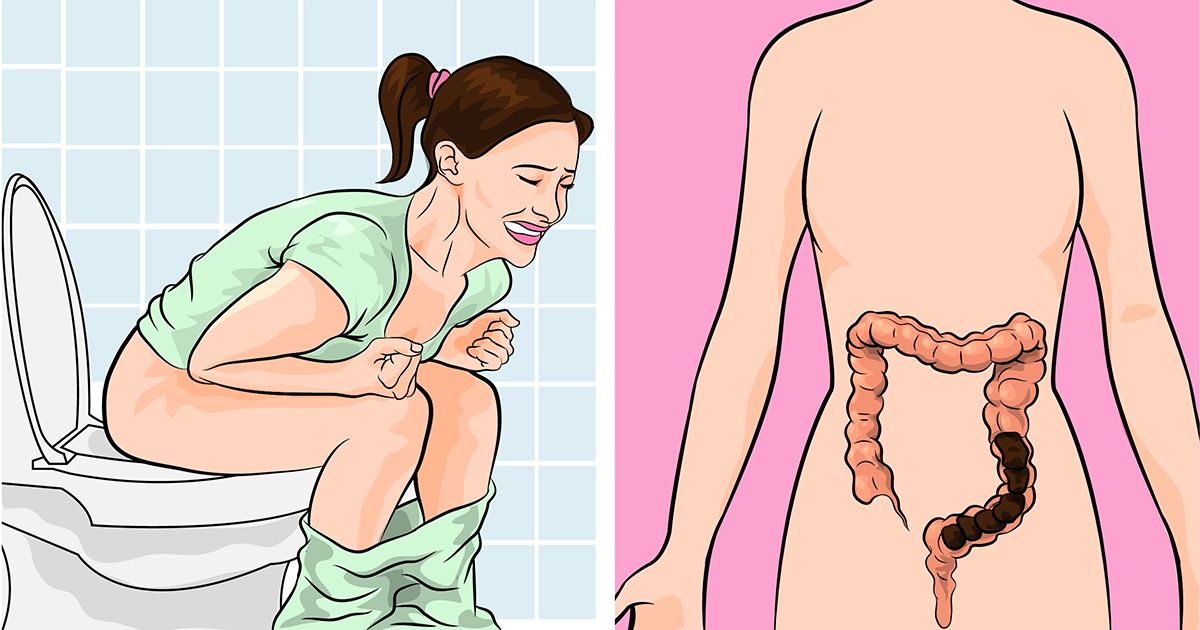
Constipation: Symptoms, Causes, Diagnosis, And Treatment Constipation is one of the mos
read more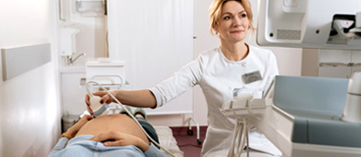
Suffering from severe bouts of intestinal pain or having swallowing problems? Go for end
read more
If you have ever searched for the “best gastroenterologist or doctor near me” or eve
read more