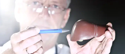

Liver Cirrhosis or Cirrhosis denotes a disease condition where the liver fails to function due to extensive damage over long period of time, involving loss of liver cells, as well as irreversible scarring of the liver. Even though at the initial stage, symptoms are negligible or nil, manifestations appear in rapid succession as the disease progresses. These include fluid buildup in the abdomen, spider-like blood vessels on the skin, swelling in lower legs and many more that are being discussed in detail next, while the patient becomes weak, loses appetite and develops yellowish skin (Jaundice). Likely to prove fatal if not treated early.
Cirrhosis symptoms may be grouped under two heads – symptoms that have resulted from failure of the liver cells and those that have occurred due to portal hypertension (secondary).
Manifestations caused due to failure of the liver cells:
Manifestations caused due to portal hypertension
Cirrhosis (liver cirrhosis) enhances resistance to blood flow and higher pressure in the portal venous system that causes portal hypertension, the effects of which are listed below.
Cirrhosis is mostly caused by excessive alcohol consumption, hepatitis B, hepatitis C, as also non-alcoholic fatty liver disorders. However, the latter is again associated with obesity, high blood pressure, high body fat and diabetes.
Some of the less common causes include autoimmune hepatitis, primary biliary cirrhosis, hemochromatosis, gall stones, as also certain types of medications. Cirrhosis is also typified by substitution of normal liver tissue with scar tissue. These result in loss of liver function.
Even though the gold standard for diagnosis of cirrhosis is liver biopsy through a percutaneous, transjugular, laparoscopic or fine-needle method, doctors currently insist on imaging as the best diagnostic approach. However, some of these approaches are outlined below.
Ultrasound is customarily used in the evaluation of cirrhosis. It may show a small and nodular liver in advanced cirrhosis along with increased echogenicity with irregular appearing areas. Other findings suggestive of cirrhosis in imaging are an enlarged caudate lobe, widening of the liver fissures and enlargement of the spleen. An enlarged spleen, which normally measures less than 11–12 cm in adults, is suggestive of cirrhosis with portal hypertension in the right clinical setting. Ultrasound may also screen for hepatocellular carcinoma, portal hypertension, and Budd-Chiari syndrome (by assessing flow in the hepatic vein).
Other tests performed in particular circumstances include abdominal CT and liver/bile duct MRI (MRCP).
Gastroscopy (endoscopic examination of the esophagus, stomach, and duodenum) is performed in patients with established cirrhosis to exclude the possibility of esophageal varices. If these are found, prophylactic local therapy may be applied (sclerotherapy or banding) and beta blocker treatment may be commenced.
Since no cure exists for biliary cirrhosis, treatment generally focuses on slowing down the progress of the disease, relieving symptoms as much as possible and preventing further complications. However, researchers are continuing to explore new drugs for treating primary biliary cirrhosis. Immunosuppressant drugs, especially methotrexate (Trexall, Rheumatrex) and colchicines (Colcrys) have been extensively tried, but their efficacy is yet to be proved.

When it comes to preserving gastrointestinal health, early diagnosis and treatment of an
read more
One of the most often occurring anorectal disorders seen in everyday practice by Indian
read more
What is Irritable Bowel Syndrome (IBS)? Irritable Bowel Syndrome (IBS) is a fairly comm
read more